The secrets of neurons revealed by water molecules
Researchers at EPFL have managed to observe the electrical activity of neurons simply by scrutinising the behaviour of the surrounding water molecules. Their simple, non-invasive method could replace the electrodes and fluorophores usually used…
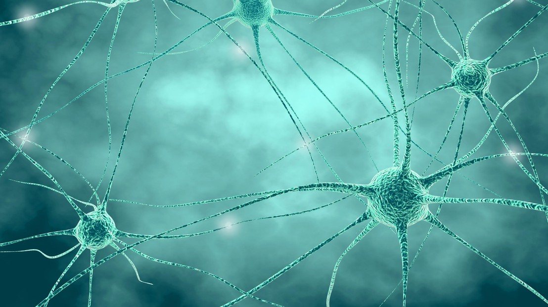
Researchers at EPFL have managed to observe the electrical activity of neurons simply by scrutinising the behaviour of the surrounding water molecules. Their simple, non-invasive method could replace the electrodes and fluorophores usually used. It makes it possible to monitor activity in a single neuron, and potentially that of an entire region of the brain.
In our brain, neurons communicate by sending electrochemical signals along axons. To trigger a signal in the form of an electrical discharge and communicate, the neuron allows ions to cross its membrane through ion channels. These ion exchanges very rapidly alter the electrical potential inside and outside the cell. This is what is known as the membrane potential.
At EPFL, the team from the Fundamental BioPhotonics Laboratory (LBP) in the School of Engineering (STI), has succeeded in observing these potential changes and the flow of ions by analysing the behaviour of the water molecules surrounding neurons. The method, which is the subject of a publication in Nature Communications, was successfully tested on mouse neurons in vitro.
Doing away with electrodes and fluorophores
The electrical activity of neurons is revealing of many phenomena in our brain. It makes it possible to tell whether a neuron is active, whether it is at rest, or whether it is responding well to a drug. At present, neurons are observed either by using fluorophores, or by placing electrodes on the area to be monitored. But fluorophores are toxic and electrodes can damage neurons.
The LBP researchers, for their part, propose monitoring electrical activity simply by observing the interactions between water molecules and the neuron membrane. “Neurons are surrounded by water molecules. These molecules change orientation according to the electrical charges present,” explains Sylvie Roke, director of the LBP. “So when the membrane potential changes, a certain number of molecules change their orientation, and we can see it.”
For their study, the researchers deliberately created changes in the membrane potential by subjecting the neurons to a large influx of potassium ions. In response, the ion channels present on the surface of the neurons -which are responsible for balancing the membrane potential- opened to let these ions in. The scientists then stopped the flow, and the neurons then released the ions that had previously entered.
To observe this activity, the researchers direct two laser beams of the same frequency at the lipid membranes of neurons immersed in water. These overlapping beams are made up of femtosecond pulses - generated thanks to the technology awarded the 2018 Nobel Prize in Physics - so that the water molecules near the membrane emit photons at double frequency, known as “second harmonic light”.
“We believe this research has both fundamental and applied implications. It may enable us to understand the mechanisms involved in the propagation of information in the brain, but it could also be of interest to pharmaceutical companies wishing to test products in vitro,” says the researcher. “We have also demonstrated that we could probe a single neuron, or several simultaneously.”.
Julia Jacobi Chair of Photomedicine - Fundamental BioPhotonics Laboratory
References
Reference: M. E. P. Didier, O. B. Tarun, P. Jourdain, P. Magistretti, and S. Roke, “Membrane water for probing neuronal membrane potentials and ionic fluxes at the single cell level”, Nature Communications
Author: Laure-Anne Pessina / Source: Mediacom

